Today we will discuss a topic that goes beyond the MRI, but that just like the MRI, allows us to “spy” inside the newborn head: the transfontanellar ultrasound.
How does it work?
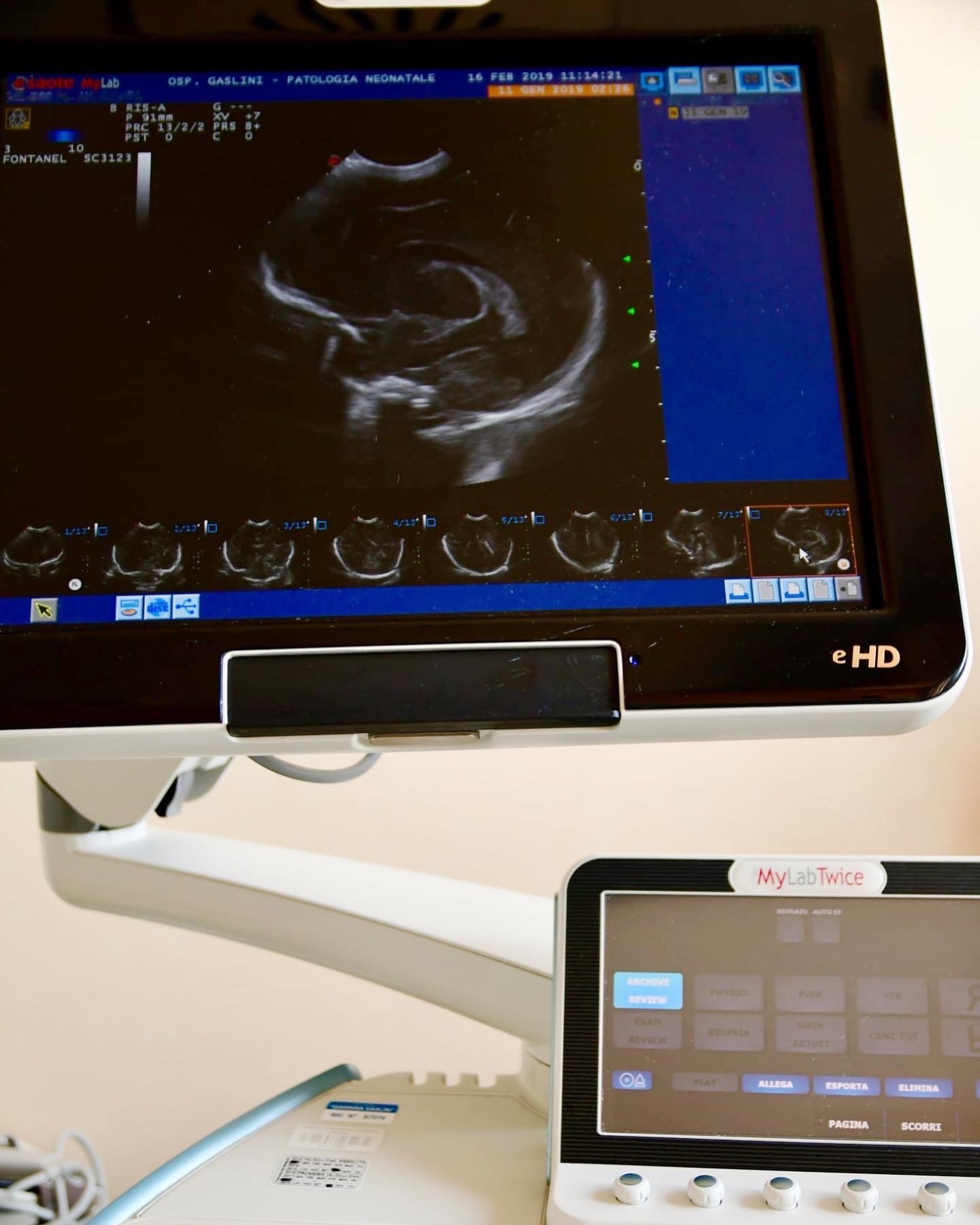
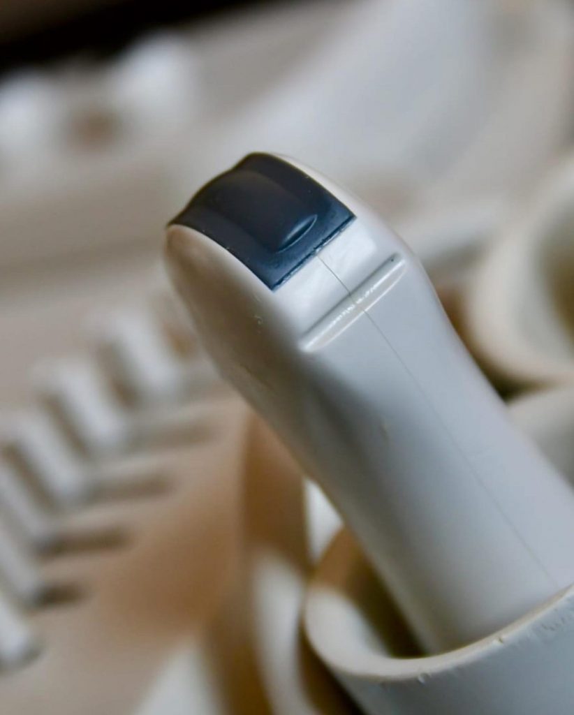
During the exam a probe sends ultrasounds inside the newborn head, which reflect on the patterns found and are transmitted back from the same probe in the form of returning echoes generating a black and white image on the computer screen.
In the newborns we leverage on the window provided by the fontanelle, joint lines between the flat bones of the newborn skull composed of fibrous tissue not yet ossified (if the bone was already formed, as in adults, we would not be able to see anything because the bone would reflect the ultrasound signal not allowing us to see underneath it). The transfontanellar ultrasound is a non-invasive test without risks that can be carried out directly in the nursery and is particularly useful for premature newborns.
It allows us to detect without using ionizing radiation and more readily than an MRI brain bleeds (the intraventricular hemorrhages we have previously discussed), damage to the periventricular white matter , hydrocephalus (build-up of fluid in the ventricles) and other rarer conditions.
Is it reliable?
Different studies have proven the excellent diagnostic reliability of the transfontanellar ultrasound in detecting matrix germinative bleedings-intraventricular hemorrhages, with the exception of first-degree brain bleeds (small bleeds) and cerebellum hemorrhages using the mastoid fontanelle.